
Evidence based overview of reflectance confocal microscopy: the 200–250µm depth limit, when it reduces facial excisions, and Rao's workflow.
200–250µm Limit: What Reflectance Confocal Microscopy Can and Can’t See
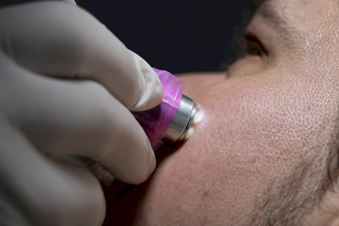
Reflectance confocal microscopy is a noninvasive imaging technique that produces near-histologic images of skin in real time, functioning as a virtual biopsy that helps dermatologists tell benign lesions from cancerous ones without a scalpel. Its primary clinical value is cutting down unnecessary excisions and pinpointing exactly where a biopsy should happen when one is needed. The tradeoff: it only sees about 200 to 250 micrometers deep, roughly the epidermis and upper papillary dermis, so it cannot stage anything invasive on its own.
TL;DR:
- Reflectance confocal microscopy cannot assess tumor invasion deeper than 200 micrometers, limiting its use for staging advanced or deep tumors.
- Different device formats exist, with bench-top systems ideal for large lesions and handheld probes suited for curved or hard-to-reach areas.
- RCM performs best as an adjunct to dermoscopy, mainly helping define lesion margins, monitor treatment response, and reduce unnecessary biopsies.
- Interpretation requires specialized training, as the imaging features vary by skin site and lesion type, with certain patterns indicating malignancy.
- Reimbursement depends on proper workflow integration, and RCM should complement, not replace, biopsy and histopathology in skin cancer diagnosis.
Table of Contents
- What Is Reflectance Confocal Microscopy (RCM)?
- How RCM Works: Optics, Contrast, and Imaging Depth
- Clinical Applications and Diagnostic Performance
- Imaging Protocol, Patient Experience, and Practice Integration
- Interpreting RCM Images: Patterns, Pitfalls, and What They Mean
- Where RCM Falls Short and Why Training Matters
- How Rao Dermatology Uses RCM in Practice
- The Gap Between What RCM Promises and How It Gets Used
- Get an Evidence-Based Skin Evaluation at Rao Dermatology
- Sources
- FAQ
What Is Reflectance Confocal Microscopy (RCM)?
RCM entered dermatology research in the 1990s and moved into routine clinical use over the following two decades as device manufacturers shrank the optics and improved image processing enough for real-time viewing. Today it sits alongside dermoscopy as a standard adjunct at academic dermatology centers and specialty skin cancer clinics, particularly for lesions on the face where a biopsy scar carries real cosmetic weight.
The technology comes in a few physical formats, and the differences matter for how a clinic actually uses it.
- Bench-top mosaic systems like the VivaScope 1500 mount a probe on an articulating arm over a glass window taped to the skin, capturing wide-field mosaics that can span a centimeter or more. This is the workhorse format for mapping large, irregular lesions such as lentigo maligna.
- Handheld devices such as the VivaScope 3000 trade field of view for maneuverability, reaching curved or tight anatomic sites, an eyelid margin, the inner ear, the nasal ala, where a bench-top probe simply cannot sit flat.
- Ex vivo systems image freshly excised tissue rather than living skin, most often during Mohs surgery to check margins in minutes instead of waiting on frozen sections.
What a clinician actually sees on screen is a grayscale horizontal section, essentially a slice parallel to the skin surface rather than the vertical cross-section a pathologist reads under a microscope. Individual images (called stacks when taken at increasing depth, mosaics when stitched side to side) reveal cell shape, nuclear brightness, and tissue architecture at a resolution close to conventional histology, minus the color and the wait for a lab result.
How RCM Works: Optics, Contrast, and Imaging Depth
RCM relies on the confocal principle: a focused laser beam illuminates one tiny point in the tissue, and a pinhole aperture in front of the detector blocks out-of-focus light from everywhere else. Only backscattered light from that exact focal point reaches the sensor, which is what makes the image so sharp compared with standard light microscopy.
The contrast comes from the tissue itself rather than any dye or stain. Melanin scatters light intensely, which is why melanocytes and pigmented cells appear bright against darker surroundings, while keratin and collagen produce their own distinct backscatter signatures that trained readers learn to distinguish by eye.
A few technical numbers define what the device can and cannot do:
- Most clinical systems use a near-infrared diode laser near 830 nanometers, a wavelength chosen because it penetrates skin reasonably well while staying safe for repeated exposure.
- Lateral resolution runs close to 1 micrometer, fine enough to make out individual nuclei and cell borders.
- Imaging depth tops out around 200 micrometers (https://pmc.ncbi.nlm.nih.gov/articles/PMC6232695/), which covers the epidermis and the papillary dermis but stops well short of the reticular dermis or subcutaneous fat.
Quick stat: That 200 to 250 micrometer ceiling is the single number every RCM report hinges on. It is deep enough to characterize a melanoma in situ or an early basal cell carcinoma, but nowhere near deep enough to rule out deep invasion, which is the whole reason RCM never replaces biopsy for anything that looks advanced.
There is also a meaningful split between how RCM is used in vivo versus ex vivo. In vivo reflectance mode, the kind used chairside on a living patient, relies purely on natural tissue backscatter. Ex vivo systems used in surgical suites often add a fluorescent dye to freshly cut tissue, which is a different confocal mode built for margin checking during surgery rather than diagnosing a lesion before it is ever cut.
Clinical Applications and Diagnostic Performance
RCM earns its place in a dermatology practice on a short list of well-studied indications, and it performs best exactly where a biopsy would otherwise leave a visible scar. Equivocal pigmented lesions, the ones that look suspicious on dermoscopy but not clearly malignant, are the classic use case: RCM lets a clinician look one layer deeper before deciding whether to cut.
Lentigo maligna mapping is arguably where RCM adds the most unique value. These lesions spread subclinically across sun-damaged facial skin with borders that are nearly impossible to judge by eye, and RCM is routinely used to define lentigo maligna margins before surgery, which matters enormously on a nose or an eyelid where every extra millimeter of excision counts.
Beyond melanoma workup, RCM has a growing role in:
- Basal cell carcinoma assessment, especially for subtle or recurrent tumors where dermoscopy alone leaves doubt.
- Amelanotic and pink tumors, lesions that lack the pigment dermoscopy depends on, where RCM’s cellular detail becomes the more useful tool.
- Squamous cell carcinoma and actinic keratosis evaluation, helping distinguish thickened sun damage from early malignant change.
- Treatment monitoring, tracking response in psoriasis, actinic keratosis undergoing field therapy, and select scalp or mucosal conditions without repeat biopsies.
Quick stat: Systematic reviews and case series of in vivo RCM report consistently high sensitivity and specificity for melanoma detection when the technique is used alongside dermoscopy rather than in isolation, reinforcing that RCM works best as a second layer of evidence, not a stand-alone verdict.
The clearest pattern across the research is where RCM changes outcomes: cosmetically sensitive sites (face, ears, genitals) and heavily sun-damaged skin where dermoscopic findings alone are ambiguous. On a trunk lesion with a classic malignant pattern, RCM rarely changes management. On a flat pigmented patch spreading across a 70 year old’s cheek, it can be the difference between one precise excision and three separate biopsies.
Imaging Protocol, Patient Experience, and Practice Integration
Getting ready for an RCM exam takes almost no preparation, which is part of its appeal for patients who already dread a biopsy visit. The MSK Cancer Center’s patient guidance advises skipping moisturizer on the area beforehand and wearing clothing that gives easy access to the site being imaged, whether that is a cheek, a shoulder, or a shin.
- Positioning. The patient sits or reclines so the target lesion is accessible and stable.
- Contact medium. A drop of immersion oil or surgical gel goes on the skin, then a small glass window or plastic ring is placed over the lesion to keep the surface flat and stable against the probe.
- Probe placement. The device rests against the window, and the clinician applies gentle, steady pressure while adjusting the angle to bring different structures into focus.
- Image capture. The system records stacks at increasing depth and, for larger lesions, stitches mosaics across the surface, a process that typically takes between a quarter to three quarters of an hour depending on lesion size and complexity.
- Cleanup and review. The gel wipes off easily, and images are reviewed either immediately or after dermatopathology correlation.
Pro Tip: If you’re heading in for an RCM exam, skip lotion or sunscreen on the area that morning. Even a thin layer of moisturizer can scatter the laser enough to blur the image and force a repeat pass.
Choosing handheld versus bench-top equipment comes down to anatomy. Bench-top mosaic systems handle large, flat lesions well, while handheld probes reach the curved and cramped sites bench-top arms cannot, though at the cost of a smaller field of view per capture.
On the business side, CMS maintains specific fee-schedule codes for RCM image acquisition and interpretation, though practices should confirm current billing rules directly with CMS and individual payers before assuming coverage, since reimbursement policy shifts and varies by plan. Adopting RCM also means budgeting staff time for training, since image quality depends heavily on operator technique.
Interpreting RCM Images: Patterns, Pitfalls, and What They Mean
Reading an RCM image well takes pattern recognition built over dozens of cases, not just knowledge of what healthy skin looks like. For melanoma and lentigo maligna, readers look for junctional atypia (irregular, enlarged cells at the dermoepidermal junction), pagetoid cells (large bright cells scattered upward into the epidermis), and general architectural disruption where the normal honeycomb pattern of keratinocytes breaks down.
Basal cell carcinoma has its own visual signature: dark silhouettes representing tumor nodules, bright elongated tumor islands, and cleft-like spaces where the tumor separates slightly from surrounding stroma, an artifact that is actually diagnostically useful rather than a flaw.
A few other patterns and caveats matter in daily practice:
- Actinic keratosis and early squamous cell carcinoma show disrupted keratinocyte patterns and atypical honeycombing without the sharp tumor islands seen in basal cell carcinoma.
- Inflammatory conditions like psoriasis show characteristic dilated papillary dermal vessels and altered epidermal architecture, useful for monitoring treatment response.
- Mucosal and acral (palm, sole) sites behave differently under RCM because the skin architecture itself differs, so readers need site-specific training rather than generalizing from facial skin.
- Thick scale, ulceration, and heavy keratin buildup all degrade image quality by scattering the laser before it reaches the target layer.
- Spitzoid lesions remain a known trap: their RCM features can overlap between benign Spitz nevi and melanoma, which is exactly the scenario where histopathology stays the final word regardless of what the images show.
Where RCM Falls Short and Why Training Matters
The 200 to 250 micrometer depth limit is RCM’s defining constraint: it cannot assess deeply invasive tumors, so it never substitutes for biopsy when a lesion looks advanced or when depth of invasion needs staging. Image quality also degrades on hyperkeratotic, ulcerated, or acral skin, and handheld probes carry their own ergonomic learning curve.
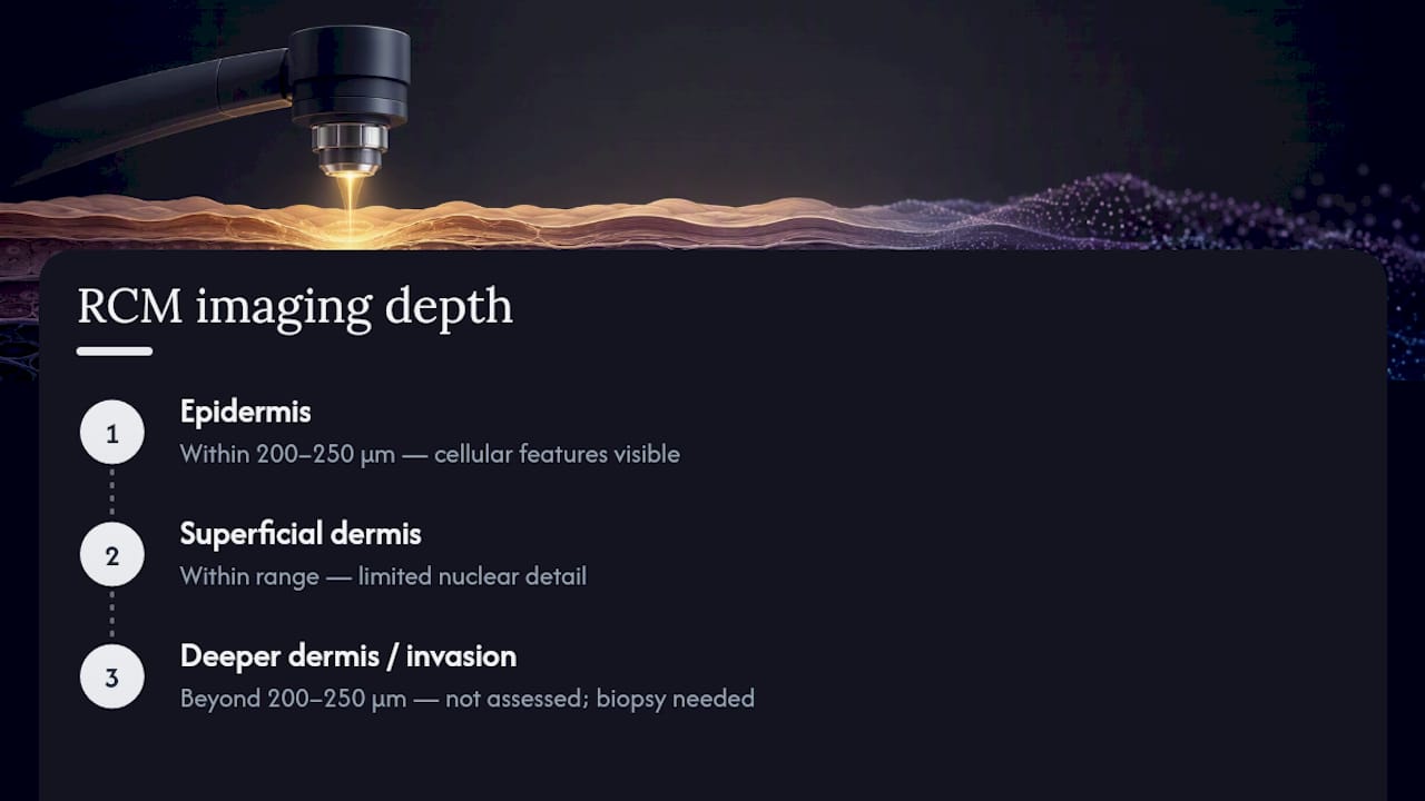
Interpretation itself demands real training. Confocal features carry lower nuclear detail than standard H&E histopathology, and interobserver variability drops meaningfully once readers gain case volume. The safest approach combines RCM with dermoscopy and keeps biopsy on the table whenever confocal findings are ambiguous or depth cannot be confirmed.
How Rao Dermatology Uses RCM in Practice
At Rao Dermatology, confocal microscopy supports evaluation of equivocal pigmented lesions, suspected lentigo maligna, and lesions on the face and other cosmetically sensitive areas where an unnecessary excision carries real cost to the patient. Findings feed directly into a broader skin cancer evaluation rather than standing alone.
The workflow pairs dermoscopy with RCM imaging, then correlates ambiguous or concerning findings with dermatopathology review before any treatment decision is finalized. Patients can expect the imaging portion of a visit to run between a quarter to three quarters of an hour depending on lesion size, similar to the process outlined in Rao Dermatology’s skin exam guide, with no downtime and no need to avoid activity afterward.
The Gap Between What RCM Promises and How It Gets Used
The biggest misconception about reflectance confocal microscopy is that it competes with biopsy. It doesn’t. The research consistently frames RCM as an add-on that improves confidence and reduces unnecessary cuts, not a replacement for tissue diagnosis, and clinics that treat it as a stand-alone verdict are misusing a genuinely good tool.
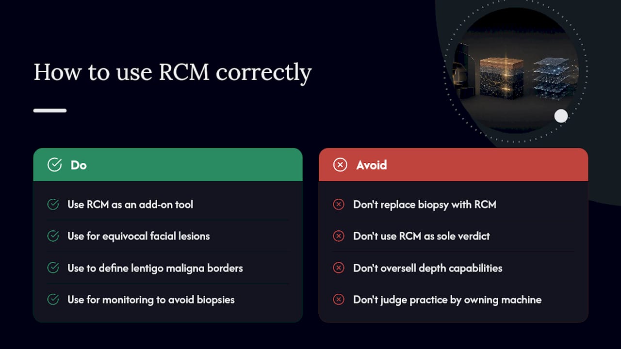
The conventional pitch oversells the technology’s reach. A 200 to 250 micrometer view sounds impressive until you realize that’s barely past the surface, useless for judging how deep a tumor actually goes. Where RCM earns its keep is narrower and more valuable than the marketing suggests: equivocal lesions on the face, lentigo maligna borders that dermoscopy can’t define, and monitoring where repeat biopsies would be excessive.
If there’s one priority for a patient or a referring clinician to take from this, it’s to ask how a given practice integrates RCM with dermatopathology rather than asking whether the practice “has” a confocal machine. The equipment matters far less than the workflow around it.
— Rao Dermatology
Get an Evidence-Based Skin Evaluation at Rao Dermatology
If you have a lesion that dermoscopy alone can’t fully explain, Rao Dermatology combines reflectance confocal microscopy with dermatopathology correlation instead of sending every questionable spot straight to the scalpel. That workflow means fewer unnecessary excisions and more precise biopsy targeting when a biopsy is genuinely needed, particularly for lesions on the face or other areas where scarring carries real weight.

Rao Dermatology’s dermatopathology and skin cancer services operate together across its California, New Jersey, and New York locations, backed by more than 25 years of clinical experience under Dr. Babar K. Rao. If a spot has you wondering whether it needs a closer look, schedule a skin evaluation through Rao Dermatology’s services page and get an answer grounded in imaging, not guesswork.
This article is general information, not a substitute for advice from a qualified doctor. Consult a qualified healthcare professional about your own circumstances before acting on anything here.
Sources
- Introduction to reflectance confocal microscopy and its use in clinical practice
- Reflectance confocal microscopy in dermatology
- Clinical Applications of In Vivo and Ex Vivo Confocal Microscopy
- Frequently Asked Questions About Reflectance Confocal Microscopy for Skin Lesions
- CMS physician fee schedule search results (RCM-related codes)
FAQ
Can Dermoscopy Alone Detect Skin Cancer?
A dermatoscope reveals surface patterns like pigment networks and vascular structures that raise or lower suspicion for cancer, but it cannot see cellular detail. Reflectance confocal microscopy adds that missing layer, which is why the two are typically used together on equivocal lesions.
What Is the Main Purpose of Confocal Microscopy in Dermatology?
Its main purpose is to provide near-histologic detail of skin lesions without cutting the skin, functioning as a virtual biopsy that helps guide whether and where to excise. It’s especially valuable for reducing unnecessary excisions on the face and other cosmetically sensitive areas.
What Are the Downsides of Using a Confocal Microscope?
The biggest limitation is imaging depth, capped around 200 to 250 micrometers, which means RCM cannot assess deeply invasive disease on its own. Image quality also suffers on hyperkeratotic, ulcerated, or acral skin, and accurate interpretation requires dedicated operator training.
What Are the Different Types of Confocal Microscopy Used in Skin Care?
The main formats are bench-top mosaic systems for wide-field imaging of larger lesions, handheld devices for curved or hard-to-reach sites, and ex vivo confocal systems used to check surgical margins on freshly excised tissue during procedures like Mohs surgery.
Does Rao Dermatology Offer Confocal Microscopy?
Confocal microscopy is used alongside dermoscopy and dermatopathology to evaluate equivocal pigmented lesions and other suspicious skin findings.
Recommended
Filed under: