
Discover how to document skin lesions accurately with our clinical guide. Use our easy EMR template for precise documentation.

Documenting a skin lesion correctly means capturing every field in a fixed, repeatable order. Here is the one-line EMR template you can paste directly into a note:
EMR Template: [Anatomic site] — [Primary morphology], [size mm × mm], [color(s)], [border], [surface/secondary changes], [configuration/distribution], [palpation], [symptoms/evolution], [dermoscopy if done], [photo IDs with scale], [assessment/plan].
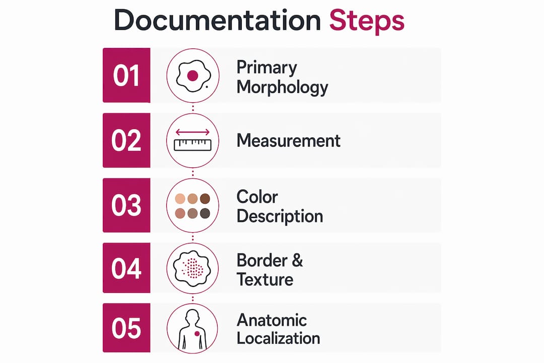
Every lesion note must include:
- Primary morphology (macule, papule, plaque, nodule, vesicle, bulla, pustule, wheal, cyst, or tumor — with the correct size-based term)
- Size in millimeters, two perpendicular axes (longest × perpendicular width)
- Color(s) with variegation noted; ABCDE criteria for any pigmented lesion
- Border (well-defined vs. ill-defined; regular vs. irregular/notched)
- Surface texture and secondary changes (smooth, scaly, crusted, eroded, ulcerated, lichenified)
- Configuration and distribution (grouped, annular, linear, dermatomal, generalized, sun-exposed)
- Palpation (firm, soft, fluctuant, mobile, fixed; depth estimate)
- Symptoms and evolution (onset, rate of change, itch, pain, bleeding)
- Dermoscopy findings if performed, using IDS consensus terminology
- Clinical photographs with scale, linked by image ID, stored in a HIPAA-compliant system
- Assessment and plan (working impression labeled separately from objective findings)
Table of Contents
- 1. Primary lesion types and the exact phrasing clinicians should use
- 2. Secondary morphology, configuration, and precise anatomical localization
- 3. How to measure lesions so the numbers mean something at follow-up
- 4. Color documentation and the ABCDE approach for pigmented lesions
- 5. Describing borders, surface texture, palpation findings, and secondary changes
- 6. Clinical photography: consent, technique, file naming, and HIPAA-compliant storage
- 7. When to use dermoscopy and how to report findings in the note
- 8. Special documentation considerations for skin of color, pediatrics, and sensitive sites
- 9. Red flags that require urgent biopsy or referral, and documentation mistakes to avoid
- 10. Ready-to-use documentation template and two example clinical notes
- Key Takeaways
- Why structured documentation is the most underrated clinical skill
- When Raodermatology can take the next step for your patient
- Useful sources
- FAQ
1. Primary lesion types and the exact phrasing clinicians should use
The foundation of any skin lesion documentation is naming the primary morphology correctly before adding any diagnostic label. Objective morphological description should precede diagnostic naming — writing “5 mm brown papule, round, smooth surface, well-defined border” is more defensible and reproducible than writing “mole” or “spot.” The Merck Manual defines the 10 mm / 1 cm threshold that separates paired lesion types.
| Primary Lesion | Size Threshold | Key Feature | Example Phrase |
|---|---|---|---|
| Macule | ≤ 10 mm | Flat, color change only | “7 mm hyperpigmented macule, brown” |
| Patch | > 10 mm | Flat, color change only | “3 cm erythematous patch, irregular border” |
| Papule | ≤ 10 mm | Elevated, solid | “4 mm pink papule, dome-shaped, smooth” |
| Plaque | > 10 mm | Elevated, flat-topped, solid | “2 cm erythematous plaque, well-defined, scaly” |
| Vesicle | ≤ 10 mm | Fluid-filled, thin-walled | “3 mm clear vesicle, tense, grouped” |
| Bulla | > 10 mm | Fluid-filled, thin-walled | “2 cm tense bulla, serous fluid” |
| Pustule | Any size | Purulent fluid | “5 mm pustule, yellow, follicular base” |
| Nodule | > 10 mm, deep | Solid, palpable depth | “1.5 cm firm nodule, dermal, mobile” |
| Tumor | greater than a typical large size | Large solid mass | “3 cm firm tumor, fixed, irregular surface” |
| Wheal | Variable | Transient, edematous | “2 cm erythematous wheal, evanescent” |
| Cyst | Variable | Enclosed sac, fluctuant | “1 cm cystic nodule, fluctuant, mobile” |
The ordering rule is non-negotiable: primary lesion type first, then size in millimeters, then color, then border, then surface. Clinicians who reverse this order — leading with color or location — produce notes that are harder to scan and easier to misinterpret during handoffs. Adopting internationally accepted controlled vocabulary from the ILDS reduces ambiguity across institutions and makes records usable for research as well as clinical care.
Pro Tip: Never use the word “lesion” alone as your primary descriptor. “Lesion” is a category, not a morphological term. Write the specific type — papule, plaque, nodule — every time.
2. Secondary morphology, configuration, and precise anatomical localization
Once you have named the primary lesion type, secondary changes and configuration tell the story of what the lesion has done over time. Scale, crust, erosion, and lichenification each point toward different diagnoses and different urgency levels.
Secondary changes to document:
- Scale: Silvery (psoriasiform), fine, adherent, or loose — note the color and adherence
- Crust: Honey-colored (impetigo), hemorrhagic, or serous
- Erosion: Superficial loss of epidermis, moist base, does not scar
- Ulceration: Full-thickness epidermal and dermal loss, may scar — document depth and base character
- Excoriation: Linear, self-induced epidermal disruption
- Lichenification: Thickened skin with accentuated markings from chronic rubbing
- Atrophy: Thinning of epidermis or dermis; note if epidermal or dermal
Configuration terms:
- Annular: Ring-shaped with central clearing (tinea, erythema migrans)
- Linear: Arranged in a line (contact dermatitis, Blaschko lines)
- Grouped/herpetiform: Clustered vesicles (herpes simplex, zoster)
- Satellite: Small lesions near a larger one (candidiasis, melanoma metastasis)
- Confluent: Lesions merging together
- Dermatomal: Following a dermatome (herpes zoster)
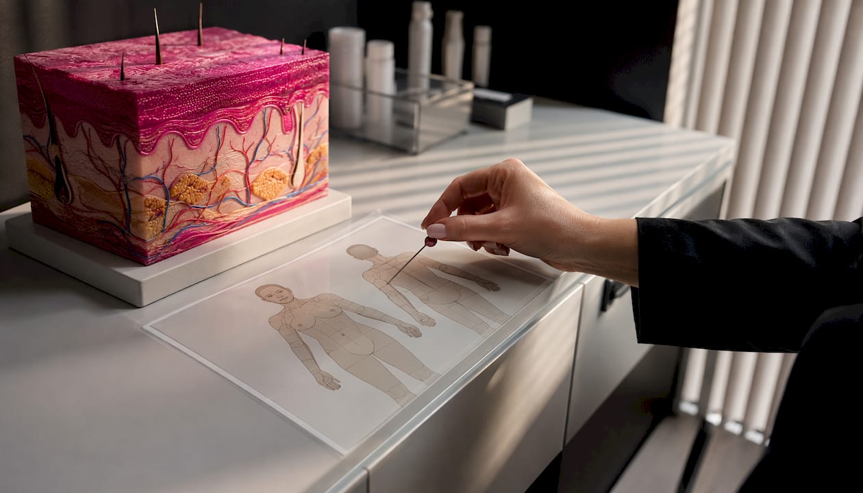
For anatomical localization, the gold standard is a body-map pin combined with a clock-face reference and a measured distance from a fixed bony landmark. A note that reads “left lateral mid-back” is not locatable at follow-up. A note that reads “left lateral mid-back, 4 cm medial to the inferior border of the left scapula, 2 o’clock position relative to the lesion center” is. Use that level of specificity every time a lesion will be monitored or biopsied. For the face, reference the canthi, nasal ala, or tragus. For the trunk, reference the umbilicus, nipple line, or iliac crest.
3. How to measure lesions so the numbers mean something at follow-up
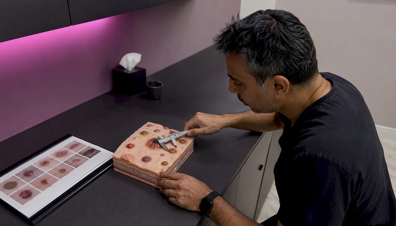
Size in millimeters is the single most important quantitative field in a lesion note. A measurement recorded as “small” or “about a centimeter” is clinically useless six months later when a different clinician is deciding whether the lesion has grown. Structured dermatology notes must record size in millimeters using two perpendicular measurements to allow longitudinal tracking.
Step-by-step measurement technique:
- Position the patient so the lesion is flat and accessible without skin tension.
- Place a millimeter ruler or digital caliper flush against the skin surface — do not measure over clothing or dressings.
- Measure the longest axis first, then the perpendicular width at the widest point.
- Record as: longest axis mm × perpendicular width mm (e.g., “14 × 9 mm”).
- Note the instrument used: “measured with flexible mm ruler” or “digital caliper.”
- For elevated lesions (nodules, cysts), estimate height in millimeters and note whether the lesion was compressed during measurement.
Recommended tools:
- Flexible millimeter ruler (disposable or sterilizable)
- Digital caliper for nodules and cysts where height matters
- Dermatoscope with built-in scale for dermoscopic close-ups
Pro Tip: Document whether the lesion was measured before or after cleansing and whether it was compressed. A fluctuant cyst measured under compression will read smaller than its true size — that discrepancy matters if you are tracking growth.
For lesions that will be biopsied, record the pre-procedure measurement in the procedure note separately from the clinical note so the pathology report can be correlated to the clinical size.
4. Color documentation and the ABCDE approach for pigmented lesions
Color is where clinical notes most often go vague. “Dark” and “pigmented” are not useful descriptors. Use these standardized color terms: pink/erythematous, red, brown, dark brown, black, violaceous, hypopigmented, depigmented (white), yellow, gray, blue. When a lesion shows more than one color, list each one and note the distribution — for example, “brown with central black focus and peripheral gray halo.”
For any melanocytic or pigmented lesion, the ABCDE criteria provide a structured framework that belongs in the note:
- A — Asymmetry: One half does not mirror the other
- B — Border: Irregular, notched, or poorly defined edges
- C — Color: Variegation — multiple shades of brown, black, red, white, or blue within a single lesion
- D — Diameter: Greater than 6 mm is a key melanoma screening threshold; note that smaller melanomas do occur, and evolution matters even for lesions under 6 mm
- E — Evolution: Any change in size, shape, color, or new symptoms (bleeding, itch, crusting)
A benign-appearing lesion note might read: “8 mm uniformly brown papule, symmetric, well-defined regular border, no variegation, stable per patient report × 5 years.” A suspicious note reads: “11 mm asymmetric macule, irregular notched border, variegated brown-black with peripheral gray, evolution: patient reports size increase over 3 months — ABCDE score positive for B, C, D, E; dermoscopy performed.”
Fitzpatrick skin type is worth recording when color interpretation depends on baseline pigmentation. In skin of color, erythema may appear violaceous or brown rather than pink, and post-inflammatory hyperpigmentation can obscure lesion borders. Note the Fitzpatrick type (I–VI) and use descriptors like “hyperpigmented relative to surrounding skin” rather than assuming a universal color baseline.
5. Describing borders, surface texture, palpation findings, and secondary changes
Border language directly affects surgical planning and referral decisions. A well-defined, regular border on a pigmented lesion supports a benign working impression; an ill-defined, notched border on the same lesion changes the urgency. Use these four border descriptors consistently:
- Well-defined: Sharp demarcation from surrounding skin
- Ill-defined: Gradual fade into surrounding skin, difficult to trace
- Regular: Smooth, even perimeter
- Irregular/notched: Scalloped, jagged, or asymmetric perimeter
Surface texture descriptors to combine with primary morphology:
- Smooth: No surface disruption
- Rough/verrucous: Warty, papillomatous surface
- Scaly: Visible scale; note adherence and color
- Crusted: Dried exudate overlying the lesion
- Erosive: Moist, superficially denuded surface
- Ulcerated: Full-thickness loss; describe base (clean, necrotic, granulating) and depth
Palpation findings to record every time:
- Firm vs. soft: Firm suggests fibrotic or infiltrative process; soft suggests lipoma or cyst
- Fluctuant: Fluid-filled; compress gently to confirm
- Mobile vs. fixed: Fixed to underlying fascia or bone raises concern for malignancy
- Depth: Superficial (epidermal/dermal) vs. deep (subcutaneous, fascial)
A complete combined descriptor might read: “1.8 cm firm, fixed nodule, ill-defined border, smooth surface, non-tender on palpation, appears to extend to deep dermis or subcutaneous plane.” That sentence gives a surgeon, pathologist, and follow-up clinician everything they need to understand the lesion without seeing the patient.
6. Clinical photography: consent, technique, file naming, and HIPAA-compliant storage
Clinical photographs without a reference scale or a keyed anatomical site are one of the most common and avoidable documentation errors. A photo that cannot be matched to a body location or compared to a follow-up image has limited medicolegal or clinical value. Photography standards require at minimum: consent documentation, date/time, anatomic site, scale, at least one overview plus one close-up, and secure HIPAA-compliant storage with the image ID linked in the note.
Photography checklist:
- Obtain and document consent before photographing (see sample phrase below)
- Overview image: full body region showing anatomic context, patient positioned consistently
- Close-up image: lesion filling 60–80% of frame, millimeter ruler in frame, consistent lighting
- Dermoscopic image: if dermoscopy performed, capture with scale and orientation marker
- Lighting: diffuse, consistent — avoid harsh flash directly on the lesion
- Background: plain, neutral (blue or green drape) to avoid distracting elements
- Focus: sharp on lesion edges, not the surrounding skin
- Orientation: mark superior/inferior or use a clock-face sticker for non-symmetric lesions
Sample consent phrase to document in the note: “Patient provided verbal consent for clinical photography of [anatomic site] for medical documentation purposes. Consent documented in chart.”
Recommended file-naming convention:
[PatientID]_[YYYYMMDD]_[AnatomicSite]_[LesionType]_[ViewType]
Example: PT12345_20260315_LeftBack_PigmentedNevus_Closeup
Store all images in a HIPAA-compliant EMR or dedicated dermatology imaging platform. Link each image to the lesion record by image ID in the clinical note: “Clinical photographs obtained — image IDs: IMG-001 (overview), IMG-002 (close-up with ruler), IMG-003 (dermoscopy) — stored in [EMR system], linked to this encounter.”
7. When to use dermoscopy and how to report findings in the note
Dermoscopy should be documented for any suspicious pigmented lesion, any lesion with reported evolution, and whenever dermoscopic evaluation meaningfully changes diagnostic confidence. The International Dermoscopy Society recommends specific analytic parameters over metaphorical terms to improve reproducibility across clinicians and institutions.
Core dermoscopic parameters to report:
- Pigment network: Typical (regular mesh) vs. atypical (irregular, broadened, or absent)
- Globules/dots: Regular distribution (benign) vs. irregular, peripheral clustering (suspicious)
- Streaks: Radial streaming or pseudopods at the periphery
- Blue-white veil: Irregular blue-white structureless area over raised portion
- Vascular patterns: Dotted, comma-shaped, irregular, or arborizing vessels
- Regression structures: White scar-like areas or peppering
A concise dermoscopy note reads: “Dermoscopy: atypical pigment network, irregular globules at periphery, focal blue-white veil — features concerning for melanoma. Dermoscopy images linked (IMG-003).”
The ILDS and IDS consensus documents are the preferred terminology sources for both neoplastic and non-neoplastic dermatoses. For non-neoplastic conditions, report vessel morphology and distribution, scale distribution, follicular findings, and other structures using the same analytic framework. Total-body photography and sequential digital dermoscopy (SDD) are best practice for high-risk patients; reference prior SDD sessions by date and image set ID in the follow-up note: “Compared to SDD session 2025-09-10 (Image Set B): lesion 3L shows 1.5 mm increase in longest axis and new peripheral globules.”
8. Special documentation considerations for skin of color, pediatrics, and sensitive sites
Standard color descriptors were developed largely on lighter skin tones, and applying them uncritically in skin of color produces inaccurate notes. Erythema in Fitzpatrick types V–VI may appear brown, violaceous, or gray rather than pink or red. Post-inflammatory hyperpigmentation can obscure lesion borders and mimic pigmented lesions. Use modifiers like “hyperpigmented relative to surrounding skin” or “violaceous plaque on dark brown skin” rather than assuming a universal color baseline.
For mucosal and genital sites, document chaperone presence and patient consent for the exam explicitly: “Exam of [site] performed with chaperone [name/role] present; patient consented verbally.” Use anatomically precise, clinical language in the chart — avoid colloquial or euphemistic phrasing that could be misread. For genital lesions, note the exact sub-site (glans, labia majora, perianal) and clock-face position.
Pediatric documentation requires parental or guardian consent for both the exam and photography. Note the child’s age, developmental stage, and whether sedation or distraction techniques were used. For congenital nevi in children, document size relative to body surface area and track growth against age-appropriate norms. If a full exam cannot be completed due to pain, movement, or refusal, document that explicitly: “Full exam of [site] limited by patient movement/pain/refusal; visible portion documented as above; remainder deferred.”
9. Red flags that require urgent biopsy or referral, and documentation mistakes to avoid
Some findings require same-day or next-available specialist contact, not a routine referral. Recognizing these in the note and documenting the action taken protects both the patient and the clinician.
Urgent red flags — document and act:
- Ulcerated, growing, irregularly pigmented lesion with any ABCDE-positive feature
- Rapid evolution over days to weeks (not months)
- Spontaneous bleeding from a pigmented or nodular lesion
- New persistent nodule in an immunosuppressed patient (transplant, HIV, chronic immunosuppressive therapy)
- Suspicious dermoscopy features (blue-white veil, atypical network, irregular vessels)
- Any lesion the clinician cannot confidently classify after full exam and dermoscopy
Urgent referral wording to paste into an EMR order: “Urgent dermatology referral requested for [anatomic site] — [primary morphology, size, ABCDE findings] — concern for [working impression]. Please evaluate within [timeframe]. Clinical photographs attached (Image IDs: [IDs]).”
Common documentation errors that make notes clinically useless or legally vulnerable:
- Using “spot,” “growth,” or “lesion” without a morphological term
- Recording size as “small,” “large,” or “about 1 cm” instead of measured millimeters
- No clinical photograph, or photograph without a ruler or anatomical context
- No anatomical landmark — “back” is not a location
- Failing to document consent for photography
- Writing a diagnosis in the objective exam field instead of a working impression
- No evolution or symptom history — a lesion with no documented timeline cannot be triaged
- Skipping dermoscopy documentation when dermoscopy was performed
For biopsy indications, a defensible note reads: “Biopsy indicated for [anatomic site] [morphology, size, ABCDE findings] — clinical concern for [working impression]. Shave/punch/excisional biopsy planned. Patient counseled on procedure, risks, and follow-up. Consent obtained and documented.”
10. Ready-to-use documentation template and two example clinical notes
The template below maps to every field discussed in this guide. Paste it into your EMR and fill in the brackets.
Full Lesion Documentation Template:
| Field | Content |
|---|---|
| Anatomic site | [Body region, clock-face, distance from landmark] |
| Primary morphology | [Macule/papule/plaque/nodule/etc.] |
| Size | [mm × mm; instrument used] |
| Color | [Descriptor(s); variegation noted] |
| Border | [Well/ill-defined; regular/irregular] |
| Surface | [Smooth/scaly/crusted/eroded/ulcerated] |
| Secondary changes | [Scale, crust, erosion, lichenification, atrophy] |
| Configuration/distribution | [Annular/linear/grouped/dermatomal/generalized] |
| Palpation | [Firm/soft/fluctuant; mobile/fixed; depth] |
| Symptoms | [Itch, pain, bleeding, tenderness] |
| Evolution | [Onset, rate of change, prior treatment] |
| Dermoscopy | [Parameters per IDS; image IDs if captured] |
| Photos | [Image IDs; overview + close-up + ruler + dermoscopy] |
| Working impression | [Differential, clearly labeled as impression] |
| Plan | [Monitor, biopsy, referral, treatment, follow-up interval] |
Example Note 1 — Stable, benign-appearing lesion:
S: 42-year-old male presents for routine skin check. Reports a lesion on the right forearm present for approximately 8 years, no change in size, color, or symptoms. No bleeding, itch, or pain.
O: Right volar forearm, 6 cm distal to the antecubital fossa — 7 × 6 mm uniformly brown papule, symmetric, well-defined regular border, smooth surface, no secondary changes, solitary. Firm, mobile, non-tender on palpation, superficial dermal depth. Dermoscopy: typical pigment network, regular globule distribution, no atypical features. Photos obtained: IMG-101 (overview), IMG-102 (close-up with ruler), IMG-103 (dermoscopy) — stored in EMR, linked to this encounter.
A: Benign-appearing melanocytic nevus. No ABCDE-positive features.
P: Reassure patient. Annual monitoring. Return sooner if evolution noted. Photos filed for longitudinal comparison.
Example Note 2 — Evolving pigmented lesion with biopsy plan:
S: 58-year-old female presents with a lesion on the left upper back, noticed approximately 6 months ago. Reports increase in size and darkening over the past 2 months. Occasional itch. No bleeding.
O: Left lateral upper back, 3 cm inferior to the inferior border of the left scapula, 2 o’clock position — 13 × 9 mm asymmetric macule-papule, ill-defined irregular notched border, variegated brown-black with peripheral gray halo, no scale or crust. Non-tender, firm, non-fluctuant, appears superficial dermal. Dermoscopy: atypical pigment network, irregular peripheral globules, focal blue-white veil, no regression structures identified. ABCDE: positive for A, B, C, D, E. Photos: IMG-201 (overview), IMG-202 (close-up with ruler), IMG-203 (dermoscopy) — stored in EMR.
A: Working impression: atypical melanocytic lesion, cannot exclude melanoma. Urgent evaluation warranted.
P: Excisional biopsy with 1–2 mm margins planned. Patient counseled on procedure, risks, and pathology follow-up timeline. Consent obtained and documented. Urgent dermatology referral placed if biopsy deferred pending specialist availability.
Photography checklist for each session:
- [ ] Consent documented in note
- [ ] Overview image (anatomic context, patient positioned consistently)
- [ ] Close-up image (ruler in frame, lesion fills 60–80% of frame)
- [ ] Dermoscopy image if performed (scale and orientation marker)
- [ ] File named per convention:
[PatientID]_[YYYYMMDD]_[Site]_[Type]_[View] - [ ] Images stored in HIPAA-compliant system and image IDs recorded in note
Key Takeaways
Systematic skin lesion documentation — primary morphology, two-axis millimeter measurement, ABCDE for pigmented lesions, precise anatomical localization, and linked clinical photographs with scale — is the minimum standard for defensible, reproducible clinical records.
| Point | Details |
|---|---|
| Use standardized morphology terms | Name the primary lesion type (papule, plaque, nodule) before any diagnostic label, following Merck Manual size thresholds. |
| Measure in millimeters, two axes | Record longest axis by perpendicular width in measurable units; note the instrument used for reproducible longitudinal comparison. |
| Apply ABCDE for pigmented lesions | Document all five criteria; diameter above a characteristic size is a key threshold, but evolution alone warrants concern in smaller lesions. |
| Photograph with scale and landmark | Every photo session needs a ruler in frame and an anatomical landmark in the note; link image IDs to the encounter record. |
| Raodermatology specialist evaluation | When documentation reveals suspicious features or uncertain diagnosis, Raodermatology offers dermoscopy-based assessment and skin cancer evaluation across California, New Jersey, and New York. |
Why structured documentation is the most underrated clinical skill
Most clinicians think of documentation as a billing requirement or a legal formality. That framing misses what a well-written lesion note actually does: it turns your clinical reasoning into a retrievable record that another clinician can act on without seeing the patient. The note is not a summary of what you saw. It is a reconstruction of the lesion that a surgeon, pathologist, or oncologist can use independently.
The real risk of vague documentation is not a malpractice claim — it is premature diagnostic closure. When a note reads “benign-appearing mole, monitor,” the next clinician has no baseline to compare against. They cannot tell whether the lesion has changed because there is no objective record of what it looked like before. Structured notes with millimeter measurements, ABCDE scoring, and linked photographs eliminate that gap. They make the chart a clinical tool, not just a record.
There is also a subtler benefit that gets overlooked: writing a structured note forces you to complete the exam. Clinicians who use templates with discrete fields — size, border, dermoscopy, evolution — are less likely to skip a field than those writing free-form narrative. The template is not bureaucracy. It is a checklist that catches the things you would otherwise assume you noticed.
Adopting standardized vocabulary from the ILDS and IDS is not about academic rigor for its own sake. It is about making your notes legible to a colleague in a different institution, a specialist reviewing a referral, or a researcher analyzing outcomes. The clinician who writes “atypical pigment network with irregular peripheral globules” is communicating something precise. The clinician who writes “funny-looking mole” is not.
When Raodermatology can take the next step for your patient

When your documentation reveals a suspicious pigmented lesion, an uncertain diagnosis after full exam and dermoscopy, or a lesion requiring excision or complex reconstruction, that is the point where specialist input changes outcomes. Raodermatology, founded by Dr. Babar K. Rao with 25+ years of dermatology experience across California, New Jersey, and New York, offers dermoscopy-based lesion assessment, skin cancer detection and treatment, and in-house dermatopathology so biopsy results are interpreted in the same clinical context as the original exam.
For referring clinicians, the dermatology patient referral checklist on the Raodermatology site walks through exactly what to include in a referral letter so the specialist receives the documentation fields this guide covers. When your note is complete and your concern is documented, the referral is straightforward. Contact Raodermatology to schedule a specialist evaluation or to discuss a complex case with the clinical team.
Useful sources
These are the primary clinical references cited throughout this guide. Consult them directly for protocol development, training, and institutional standards.
- Merck Manual Professional — Description of Skin Lesions: The standard reference for primary lesion morphology terminology and size thresholds separating paired lesion types. Use for training and terminology disputes.
- International League of Dermatological Societies (ILDS): The global consensus body for dermatological terminology. Consult for controlled vocabulary, especially when documenting for research or cross-institutional records.
- International Dermoscopy Society — Consensus on Dermoscopic Parameters: The IDS consensus document on standardized dermoscopic terminology for both neoplastic and non-neoplastic dermatoses. The preferred source for dermoscopy reporting vocabulary.
- PMC — Standards for Documenting Skin Lesions and Photography: Peer-reviewed guidance on baseline measurements, serial photography, and the requirement for scale and anatomical localization in clinical images.
- National Cancer Institute — Melanoma Screening and ABCDE Criteria: The primary U.S. government source for ABCDE melanoma screening criteria and the 6 mm diameter threshold.
- NCBI Bookshelf — Clinical Methods: The Skin and Appendages: Classic clinical reference supporting objective morphological description before diagnostic labeling and the concept of working impression as a separate field.
- Raodermatology — Dermatopathology Service: Raodermatology’s in-house dermatopathology capabilities, relevant to biopsy workflow and pathology correlation documentation.
FAQ
How do you medically describe a skin lesion?
Use a fixed order: primary morphology (macule, papule, plaque, nodule), size in millimeters (two perpendicular axes), color(s), border (well-defined vs. ill-defined, regular vs. irregular), surface texture, secondary changes, configuration, and anatomical location referenced to a fixed landmark. For pigmented lesions, add ABCDE criteria.
How do you document skin findings on a physical exam?
Record each lesion using the standardized template: anatomic site with landmark, primary morphology, size in mm × mm, color, border, surface, secondary changes, configuration, palpation findings, symptoms, evolution, dermoscopy if performed, and linked photo IDs. Keep the objective exam separate from the working impression field.
How do you describe the distribution of skin lesions?
State whether the distribution is localized or generalized, then specify the pattern: dermatomal (following a nerve root), sun-exposed (face, dorsal hands, V of neck), flexural, extensor, intertriginous, or mucosal. For grouped or annular configurations, name the configuration term and give the anatomical region with a landmark reference.
What are the most common documentation errors in dermatology notes?
The most frequent errors are using vague terms like “spot” or “growth” instead of a morphological descriptor, recording size without millimeter measurements, omitting a clinical photograph or including one without a ruler, and failing to document an anatomical landmark precise enough to relocate the lesion at follow-up.
When should a skin lesion be referred to a specialist like Raodermatology?
Refer when a lesion has ABCDE-positive features, shows rapid evolution, bleeds spontaneously, cannot be confidently classified after exam and dermoscopy, or requires excision or complex reconstruction. Raodermatology offers dermoscopy-based assessment and skin cancer evaluation across California, New Jersey, and New York.
Recommended
Filed under: