
Discover what to expect in your comprehensive skin exam guide. Learn how early detection can protect your skin and health.
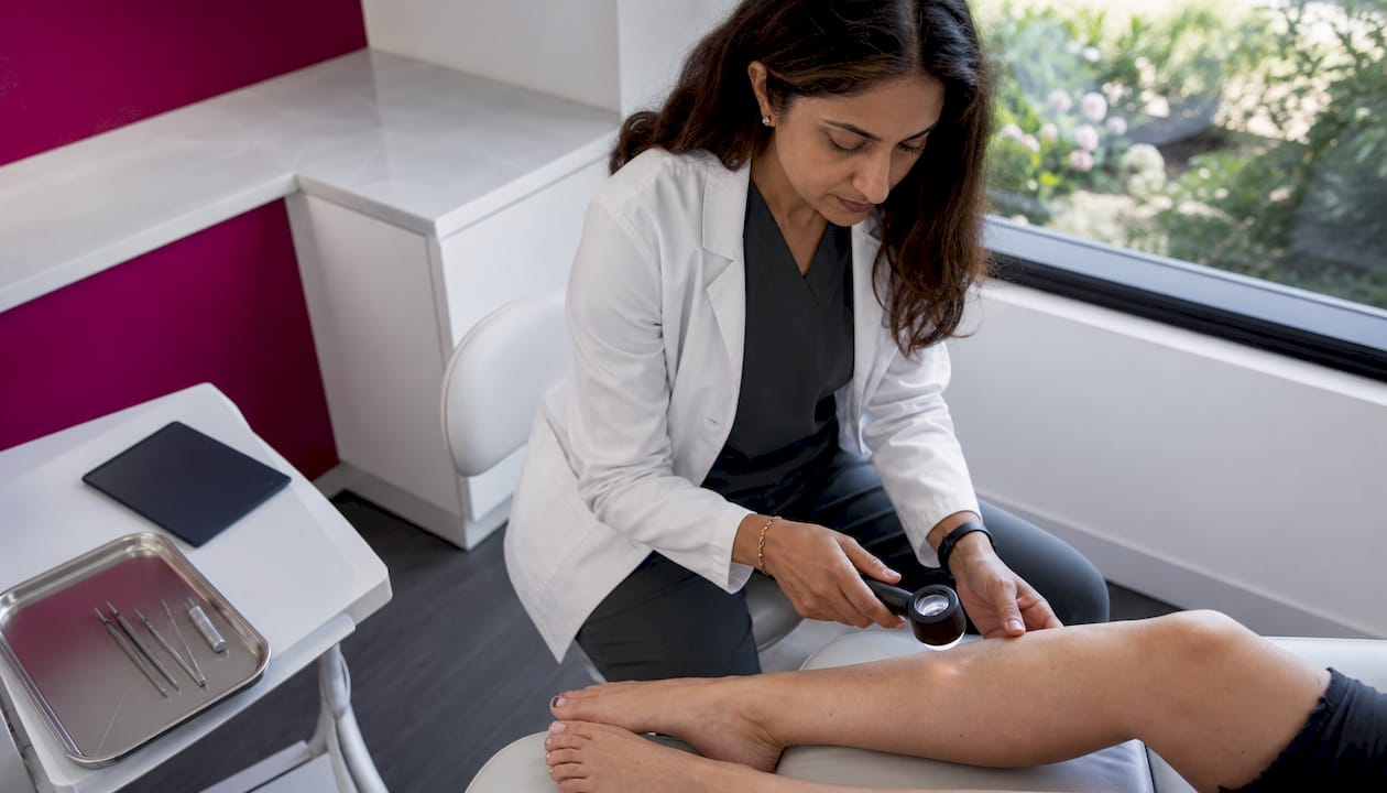
A comprehensive skin exam is a full visual inspection of your body’s skin, performed by a dermatologist to detect early signs of skin cancer and other conditions. The clinical term is a “full body skin assessment,” and it covers every surface area from your scalp to the soles of your feet. 1 in 5 Americans will develop skin cancer in their lifetime. That number makes routine screening one of the most impactful preventive steps you can take. When caught early, basal cell carcinoma (BCC) and squamous cell carcinoma (SCC) carry a cure rate near 95%. Early detection is not just reassuring. It is the difference between a minor procedure and a major one.
What does a comprehensive skin exam actually involve?
A comprehensive skin exam is a systematic, head-to-toe visual check performed by a licensed dermatologist or trained clinician. The goal is to identify any lesions, moles, or spots that look abnormal before they progress. This is not the same as a quick glance at a rash during a general physical. A full body skin assessment is methodical, thorough, and covers areas most people never think to check on their own.
The exam includes your scalp, ears, lips, eyelids, fingernails, toenails, the spaces between your toes, and, with your consent, the genital area. These are critical hidden areas that self-exams routinely miss. Melanoma found under a toenail or behind an ear is just as dangerous as one found on your back. Skipping those zones defeats the purpose of a thorough skin health evaluation.
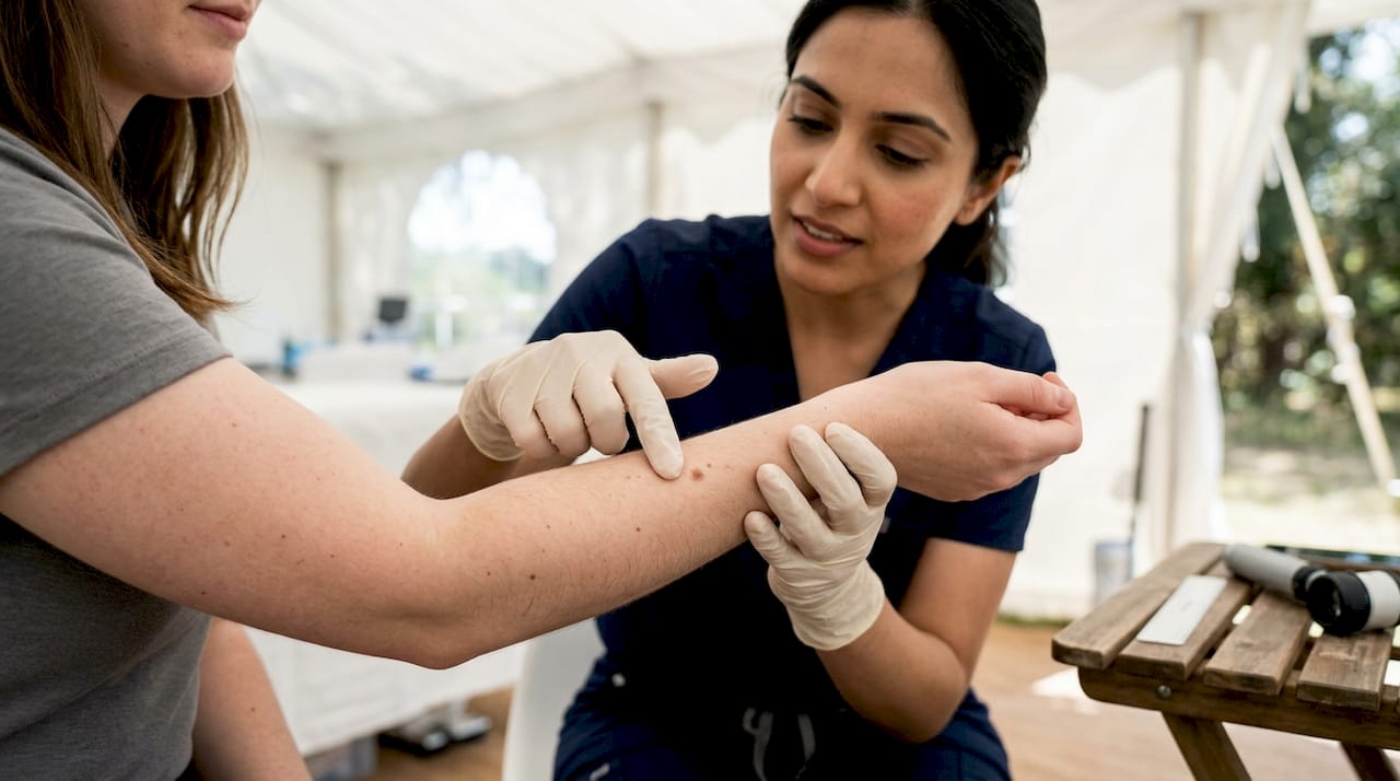
Raodermatology, founded by Dr. Babar K. Rao with over 25 years of clinical experience across California, New Jersey, and New York, performs these exams using the latest diagnostic tools, including dermatoscopy, to reduce unnecessary biopsies and improve accuracy.
How to prepare for your skin exam
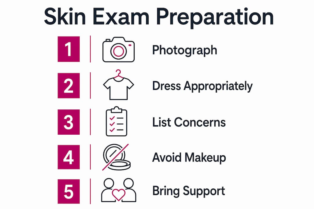
Preparation directly affects how thorough your exam can be. A dermatologist cannot evaluate what they cannot see clearly. Follow this skin check checklist before your appointment.
- Remove nail polish from fingers and toes. Nail beds are a common site for melanoma, and polish blocks the view entirely.
- Skip makeup and lotions. Skin must be clean and product-free so the clinician can assess true color and texture.
- Wear loose, easy-to-remove clothing. This speeds up the exam and reduces awkwardness when undressing.
- Do not shave or wax suspicious spots. Shaving over a suspicious mole can cause irritation or bleeding that obscures the dermatologist’s assessment.
- Compile your medical and family history. A personal or family history of melanoma, BCC, or SCC changes how aggressively your clinician monitors certain lesions.
- Document suspicious spots with photos. Taking close-up photos of changing moles days or weeks before your appointment gives your dermatologist a comparison baseline.
Pro Tip: Use your phone’s portrait mode to photograph any mole that has changed recently. Include a ruler or coin in the frame for scale. This simple step gives your dermatologist measurable data, not just your memory.
For a full preparation walkthrough, Raodermatology’s appointment prep guide covers every step in detail.
What happens during a full body skin assessment, step by step
Knowing what to expect removes most of the anxiety around this exam. A typical appointment runs 10–25 minutes and is completely painless unless a biopsy is needed.
- Check-in and history review. The clinician reviews your medical history, any medications, and your list of concerns or suspicious spots.
- Gown up. You change into a gown. Privacy is maintained throughout. The clinician exposes one area at a time, so you are never fully uncovered.
- Head-to-toe visual scan. The dermatologist works systematically, starting at the scalp and moving downward. They check your face, neck, chest, back, arms, hands, abdomen, legs, and feet.
- Dermatoscope examination. For any lesion that looks unusual, the clinician uses a dermatoscope, a handheld magnifying device with polarized light, to examine the structure beneath the skin’s surface. This tool reduces unnecessary biopsies by giving the clinician a clearer picture before cutting.
- Photography of suspicious lesions. The clinician may photograph spots they want to monitor over time.
- Biopsy if needed. If a lesion looks suspicious, a small tissue sample is taken under local anesthetic. The sample goes to a lab for analysis. Raodermatology’s dermatopathology services handle this analysis in-house, which speeds up results.
- Discussion and next steps. The dermatologist explains findings, answers questions, and outlines a follow-up plan.
Pro Tip: You can ask for a slower pace at any point. You can also bring a support person into the room. These are standard accommodations, not special requests. Speak up if you feel rushed.
What do common findings mean, and what happens next?
Most patients leave a skin exam with a clean bill of health or minor monitoring instructions. Understanding the range of possible findings helps you process results calmly.
Normal findings include benign moles (called nevi), seborrheic keratoses (rough, waxy brown spots that are harmless), and cherry angiomas (small red dots from dilated blood vessels). These require no treatment.
Suspicious findings are evaluated using the ABCDE rule:
- A: Asymmetry. One half of the mole does not match the other.
- B: Border. Edges are ragged, notched, or blurred.
- C: Color. Multiple shades of brown, black, red, white, or blue within one spot.
- D: Diameter. Larger than 6 millimeters (about the size of a pencil eraser).
- E: Evolving. Any mole that is changing in size, shape, color, or behavior.
A related concept is the “ugly duckling sign.” A mole that looks distinctly different from all your other moles warrants attention, even if it does not tick every ABCDE box. This is a practical tool for both clinicians and patients doing self-checks.
| Finding type | Typical next step |
|---|---|
| Benign mole or keratosis | Monitor annually or as directed |
| Atypical mole (dysplastic nevus) | Biopsy or close monitoring every 3–6 months |
| Suspicious lesion (possible BCC or SCC) | Biopsy within days to weeks |
| Suspected melanoma | Urgent biopsy and staging workup |
Exam frequency is tailored to your risk profile. Patients with a history of melanoma, a family history of skin cancer, or significant sun damage may need exams every 3–6 months. Most low-risk adults benefit from an annual check. Raodermatology’s skin cancer screening guide explains how to determine the right schedule for your situation.
How to perform skin self-exams between professional visits
Monthly self-exams are the best way to catch changes between professional screenings. They do not replace a dermatologist’s eye, but they give you a consistent baseline for your own skin.
What you need:
- A full-length mirror
- A handheld mirror for hard-to-see areas
- Bright, natural lighting
- A body map or photo log to track spots over time
Where to check:
- Face, scalp (use a comb or hair dryer to part hair), ears, and neck
- Chest, abdomen, and both sides of your arms
- Backs of your hands, between fingers, and under nails
- Back and buttocks (use the handheld mirror or ask someone to help)
- Legs, feet, soles, and between toes
Photographing and tracking suspicious spots between visits gives your dermatologist measurable evidence of change. A spot that looks the same in 12 photos over a year is far less concerning than one that shifts in three. Some patients use remote health monitoring tools from services like PRYM Wellness to organize their health tracking between clinical visits.
Seek professional evaluation promptly if a spot bleeds without injury, itches persistently, or changes noticeably within weeks. These are not wait-and-see situations. Raodermatology’s at-home skin check guide walks through the full self-exam process with visual guidance.
Key Takeaways
A comprehensive skin exam, performed annually and supplemented by monthly self-checks, is the most reliable way to detect skin cancer before it becomes life-threatening.
| Point | Details |
|---|---|
| Early detection saves lives | BCC and SCC caught early carry a cure rate near 95%, making routine exams worth the time. |
| Preparation matters | Arrive with clean skin, no nail polish, and a photo log of any changing spots. |
| The ABCDE rule guides evaluation | Asymmetry, border, color, diameter, and evolution are the five warning signs every patient should know. |
| Self-exams fill the gap | Monthly checks with a mirror and photo log help you catch changes between professional visits. |
| Frequency depends on risk | High-risk patients may need exams every 3–6 months; most low-risk adults benefit from an annual screening. |
Why I think most people wait too long for their first skin exam
The most common reason patients give for skipping a full body skin assessment is discomfort. Not pain. Discomfort. The idea of standing in a gown while someone examines every inch of your skin feels vulnerable. I understand that completely. But here is what I have seen over years of working in this field: the patients who feel awkward for 15 minutes are the ones who avoid a surgery, a scar, or worse.
The exam itself is far less invasive than people expect. Dermatologists do this dozens of times a day. They are looking at your skin, not judging it. And the privacy protocols, one area exposed at a time, blankets available, support persons welcome, exist precisely because patient comfort is part of good clinical practice.
What I tell patients who are nervous: tell your provider before the exam starts. Say “I’m anxious about this” or “I’d like to go slowly.” That single sentence changes the entire dynamic. You are in control of the pace. The dermatologist is there to help you, not rush you.
The other thing worth saying plainly: catching a melanoma at stage 1 versus stage 3 is not a small difference. It is often the difference between a minor excision and chemotherapy. No amount of pre-exam discomfort outweighs that outcome. Schedule the exam. You will leave wondering why you waited.
— Krunal
Raodermatology’s approach to skin cancer screening
Raodermatology brings over 25 years of clinical expertise to every skin cancer screening, with locations across California, New Jersey, and New York. The practice uses dermatoscopy and in-house dermatopathology to give patients fast, accurate results without unnecessary referrals.

Every exam at Raodermatology is conducted with patient comfort as a priority. Clinicians take time to explain findings clearly and build a monitoring plan tailored to your risk profile. Whether you are coming in for your first full body skin assessment or following up on a suspicious lesion, the team is trained to make the process straightforward. Visit the skin cancer services page to learn more or schedule your exam today.
FAQ
How long does a full body skin exam take?
A comprehensive skin exam typically takes 10–25 minutes. The appointment may run longer if the dermatologist finds multiple areas that need closer evaluation or if a biopsy is performed.
Do I need to remove all my clothes for a skin exam?
You wear a gown, and the clinician exposes one area at a time throughout the exam. Privacy is maintained at every step, and you can request a slower pace or have a support person present.
How often should I get a professional skin exam?
Exam frequency depends on your personal risk factors. Most low-risk adults benefit from an annual screening, while patients with a history of melanoma or significant sun damage may need checks every 3–6 months.
What is the ABCDE rule in skin cancer screening?
The ABCDE rule stands for Asymmetry, Border, Color, Diameter, and Evolving. These five criteria help dermatologists and patients identify moles or spots that may need further evaluation or biopsy.
Can I do a skin self-exam at home?
Monthly self-exams using a full-length mirror, a handheld mirror, and good lighting are a reliable way to track changes between professional visits. They supplement but do not replace a dermatologist’s full body skin assessment.
Recommended
- Step by Step Skin Cancer Screening: Your Full Guide | Rao Dermatology
- Your step-by-step cosmetic dermatology consultation guide | Rao Dermatology
- Essential Guide to Skin Self-Examination: What to Look for Between Dermatology Visits | Rao Dermatology
- Annual Skin Cancer Screening: Your Complete Guide to Dermatology Checkups | Rao Dermatology
Filed under: