
Discover why early skin cancer detection is important for survival. Learn how timely check-ups can save lives and your skin health.
Early skin cancer detection is defined as identifying malignant or pre-malignant skin lesions before they invade deeper tissue or spread to lymph nodes and distant organs. The survival difference between catching melanoma early versus late is not marginal. Localized melanoma exceeds 99% treatment success, while metastatic melanoma drops to roughly 30%. That gap represents lives, not statistics. Understanding why early detection matters, and what tools and habits actually work, is the most direct path to protecting your skin health in 2026.
Why early skin cancer detection is important for treatment outcomes
The biology of skin cancer explains why timing is everything. When a melanoma or basal cell carcinoma is caught at the localized stage, it has not yet penetrated the deeper dermis or entered the lymphatic system. Treatment at this point typically means a minor surgical excision with clean margins, minimal recovery, and no need for systemic therapy. Wait until the tumor thickens or spreads, and the treatment picture changes completely.
The Breslow index measures tumor thickness in millimeters and directly predicts prognosis. A melanoma under 1mm thick carries a dramatically better outlook than one exceeding 4mm. Thickness increases as time passes without detection, which is why every month of delay carries real clinical weight. Less invasive treatments become available when lesions are caught early, preserving surrounding tissue and protecting quality of life in ways that late-stage treatment simply cannot.
The advantages of early skin cancer detection go beyond survival rates. Patients diagnosed early avoid chemotherapy, immunotherapy, and radiation in most cases. They retain more normal skin architecture, face fewer surgical complications, and return to daily life faster. For non-melanoma skin cancers like squamous cell carcinoma, early detection often means the difference between a simple in-office procedure and a reconstructive surgery.
- Stage matters more than type. Even aggressive melanoma subtypes respond well to excision when caught at stage I.
- Tumor thickness is controllable. Regular screening keeps Breslow measurements low by catching lesions before they grow.
- Treatment complexity scales with delay. Each stage of progression adds treatment modalities, side effects, and cost.
- Quality of life is a measurable outcome. Early-stage patients report significantly better functional and psychological outcomes post-treatment.
Pro Tip: If you notice a mole changing in color, border, or size over four to six weeks, do not wait for your annual appointment. Schedule an evaluation immediately. The ABCDE rule (Asymmetry, Border, Color, Diameter, Evolution) is your first filter.
How do self-exams and professional screenings work together?
Self-examination and professional screening are not competing strategies. They are complementary layers of a detection system, and both are necessary. Relying on only one leaves gaps that the other fills.

76% of early-stage melanomas are first identified by patients themselves, not clinicians. This means your monthly self-exam is not a backup plan. It is the primary surveillance tool for most people between annual appointments. The catch is that self-exams have real limitations: you cannot see your own scalp, back, or the skin between your toes without deliberate effort and a mirror system.
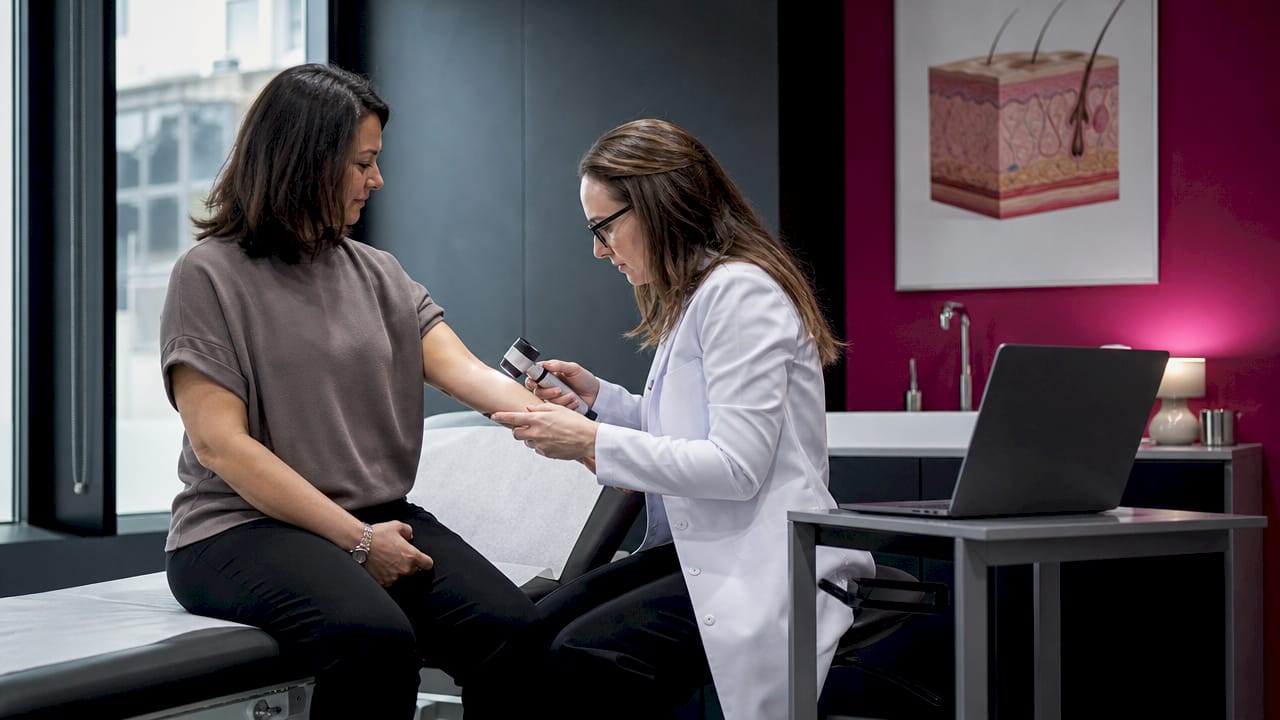
Professional full-body skin exams close those gaps. A dermatologist using dermoscopy, a technique that uses polarized light to visualize subsurface skin structures, can distinguish a benign mole from an early melanoma with far greater accuracy than the naked eye. Dermoscopy avoids unnecessary biopsies by giving clinicians a clearer picture before cutting. A routine professional screening takes about 15 minutes and covers areas you simply cannot examine yourself.
Here is how to structure an effective monthly self-exam:
- Use a full-length mirror and a hand mirror. Check your front, back, and sides in good lighting before examining harder-to-see areas.
- Examine your scalp. Use a comb or hair dryer to part your hair in sections. Ask a partner to help if needed.
- Check between your toes and the soles of your feet. Skin cancer develops in overlooked areas like these more often than most people expect.
- Document what you find. Photograph any moles or spots you want to track. Date the photos and compare them monthly.
- Know your baseline. New lesions or changes to existing ones are the signal to act, not the presence of moles themselves.
The U.S. Preventive Services Task Force currently issues an “I” (insufficient evidence) recommendation on routine clinician screening for asymptomatic adults. Self-exams remain vital regardless of that policy position, particularly for individuals with a personal or family history of skin cancer, fair skin, or significant sun exposure history. You can learn the full technique through Raodermatology’s monthly self-exam guide.
Pro Tip: Smartphone apps marketed for mole analysis are not a substitute for clinical evaluation. As of 2026, app-based detection remains unreliable. Use photos to track changes over time, but always bring findings to a dermatologist.
What advanced technologies improve early skin cancer diagnosis?
Beyond the standard visual exam, dermatology has developed a set of imaging technologies that make early diagnosis more accurate and less invasive. These tools are particularly valuable for high-risk patients who need more than an annual naked-eye check.
| Technology | Primary benefit | Best suited for |
|---|---|---|
| Dermoscopy | Subsurface visualization without biopsy | All patients, standard of care |
| Digital mole mapping | Tracks lesion changes over time | High-risk patients with many moles |
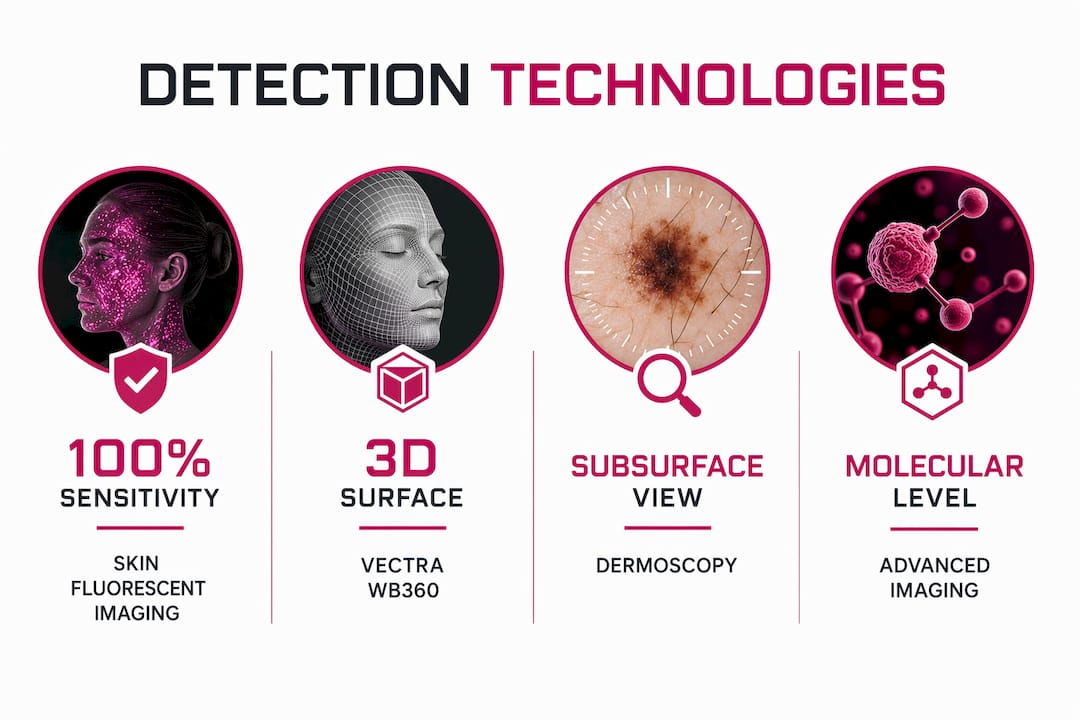
| VECTRA WB360 | Full-body 3D skin mapping in one scan | High-risk patients needing comprehensive monitoring |
| Skin Fluorescent Imaging (SFI) | Molecular-level melanoma detection | Ambiguous lesions requiring high accuracy |
VECTRA WB360 captures a detailed three-dimensional map of the entire body surface, allowing dermatologists to track subtle changes in moles and lesions across visits. For patients with dozens of atypical moles, this technology removes the guesswork from monitoring. Digital mole mapping extends this capability by creating a longitudinal record that flags new or evolving lesions that might otherwise be missed between appointments.
Molecular imaging represents the frontier of early detection. Skin Fluorescent Imaging achieves 100% sensitivity and 95.7% specificity for melanoma detection. Those numbers mean fewer false negatives and fewer unnecessary biopsies, which matters both clinically and practically. Patients avoid the anxiety and cost of biopsies that turn out to be benign, while genuinely suspicious lesions get flagged with greater confidence. You can read more about how dermoscopy works and why it remains the clinical gold standard.
High-risk individuals gain the most from combining dermoscopy with digital mole mapping and advanced imaging. If you have a history of melanoma, more than 50 moles, a family history of skin cancer, or significant cumulative sun damage, ask your dermatologist whether these tools are appropriate for your monitoring plan.
What practical steps protect you through early detection?
Knowing the importance of skin cancer screening is only useful if it translates into consistent habits. The following steps give you a structured approach to protecting yourself.
- Schedule an annual full-body skin exam. If you are high-risk, ask about twice-yearly appointments. Annual exams catch what self-exams miss and give your dermatologist a baseline for comparison.
- Perform a monthly self-exam on the same date each month. Consistency matters more than perfection. A regular schedule builds familiarity with your own skin, making changes easier to spot.
- Practice sun safety year-round. Broad-spectrum SPF 30 or higher, protective clothing, and avoiding peak UV hours between 10 a.m. and 4 p.m. reduce your cumulative risk. Preventing skin cancer and detecting it early are not separate strategies. They work together.
- Keep a photo log of your moles. A simple folder on your phone with dated images gives you and your dermatologist a visual history. This is especially useful for moles in hard-to-see locations.
- Act on changes promptly. Any mole that bleeds, itches, crusts, or changes within four to six weeks warrants a professional evaluation. Do not wait for your scheduled annual visit.
The role of dermatology in cancer detection extends beyond the exam room. A good dermatologist educates you on your personal risk profile, tailors screening frequency to your history, and uses the right technology for your situation. Understanding that relationship helps you advocate for the level of care you actually need. Raodermatology’s overview of the dermatologist’s role in skin cancer explains what to expect from that partnership.
Key takeaways
Early skin cancer detection is the single most effective intervention available, converting a potentially fatal diagnosis into a manageable, often curable condition through timely, minimally invasive treatment.
| Point | Details |
|---|---|
| Survival gap is dramatic | Localized melanoma exceeds 99% treatment success; metastatic melanoma drops to roughly 30%. |
| Self-exams drive early finds | Patients identify 76% of early melanomas themselves, making monthly checks non-negotiable. |
| Technology reduces biopsy burden | Dermoscopy and SFI distinguish benign from malignant lesions with high accuracy before cutting. |
| High-risk patients need more | Digital mole mapping and VECTRA WB360 provide longitudinal tracking beyond standard exams. |
| Prevention and detection work together | Sun safety reduces risk while regular screenings catch what prevention alone cannot stop. |
What I’ve seen change when patients stop waiting
I have reviewed enough case histories to say this plainly: the patients who do worst are not the ones with the most aggressive tumors. They are the ones who waited. A mole that looked “the same” for years, a spot dismissed as a sun spot, a lesion on the scalp that went unnoticed because no one ever looked. These are the cases that arrive at stage III or IV, and they are almost always preventable with earlier action.
What strikes me most about the current moment in dermatology is that the tools have never been better. VECTRA WB360, digital mole mapping, SFI, and high-resolution dermoscopy give clinicians a level of diagnostic precision that was unavailable even five years ago. Yet the gap between what technology can do and what patients actually do remains wide. Most people still do not perform monthly self-exams. Many high-risk individuals go years between professional screenings.
The uncomfortable truth is that skin cancer awareness campaigns have not closed that gap. What does close it is a patient who understands the specific survival numbers, knows their own risk profile, and has a dermatologist they see regularly. Education without action is just information. The patients I see who fare best are the ones who treat their annual skin exam the same way they treat a dental cleaning: non-negotiable, scheduled in advance, and never skipped because nothing looks wrong. Nothing looking wrong is exactly when you want to go.
— Krunal
Protect your skin with Raodermatology’s detection services
Raodermatology offers skin cancer screening and treatment across locations in California, New Jersey, and New York, backed by Dr. Babar K. Rao’s 25+ years of clinical expertise. The practice uses dermoscopy, digital mole mapping, and advanced imaging technologies to catch skin cancers at their earliest and most treatable stages. Whether you need a baseline full-body exam, high-risk monitoring, or evaluation of a suspicious lesion, Raodermatology’s team provides personalized care built around your specific risk profile. Schedule your skin cancer screening today and get the clarity that comes from a thorough, technology-supported examination by specialists who do this every day.
FAQ
What is the survival rate difference between early and late skin cancer?
Localized melanoma has a treatment success rate exceeding 99%, while metastatic melanoma survival falls to approximately 30%. This gap makes the timing of detection the single most important factor in skin cancer outcomes.
How often should you get a professional skin cancer screening?
Most adults benefit from an annual full-body skin exam with a dermatologist. High-risk individuals, including those with a personal or family history of melanoma, more than 50 moles, or significant sun damage, may need screenings every six months.
Can smartphone apps reliably detect skin cancer?
No. As of 2026, smartphone app-based mole analysis remains insufficient for reliable detection. Clinician examination using dermoscopy is the gold standard, and apps should only be used to photograph and track changes over time.
What are the early signs of skin cancer to watch for?
The ABCDE rule covers the key warning signs: Asymmetry, irregular Border, multiple Colors, Diameter larger than 6mm, and Evolution or change over time. Any lesion that bleeds, itches, or crusts without healing also warrants prompt evaluation.
Does early detection of skin cancer reduce the need for aggressive treatment?
Yes. Early-stage skin cancers are typically treated with surgical excision alone, avoiding chemotherapy, radiation, or immunotherapy. Catching cancer before it thickens or spreads preserves tissue, reduces recovery time, and significantly improves quality of life.
Recommended
- What Is Dermatoscopy? Guide to Early Skin Cancer Detection | Rao Dermatology
- Annual Skin Cancer Screening: Why You Need One Every Year | Rao Dermatology
- Role of dermatologists in skin cancer: A guide for patients | Rao Dermatology
- Annual Skin Cancer Screening: Your Complete Guide to Dermatology Checkups | Rao Dermatology
Filed under: