
Clinic backed guide to suspicious mole signs, photo logging every 4–12 weeks, and biopsy basics from a dermatology clinic.
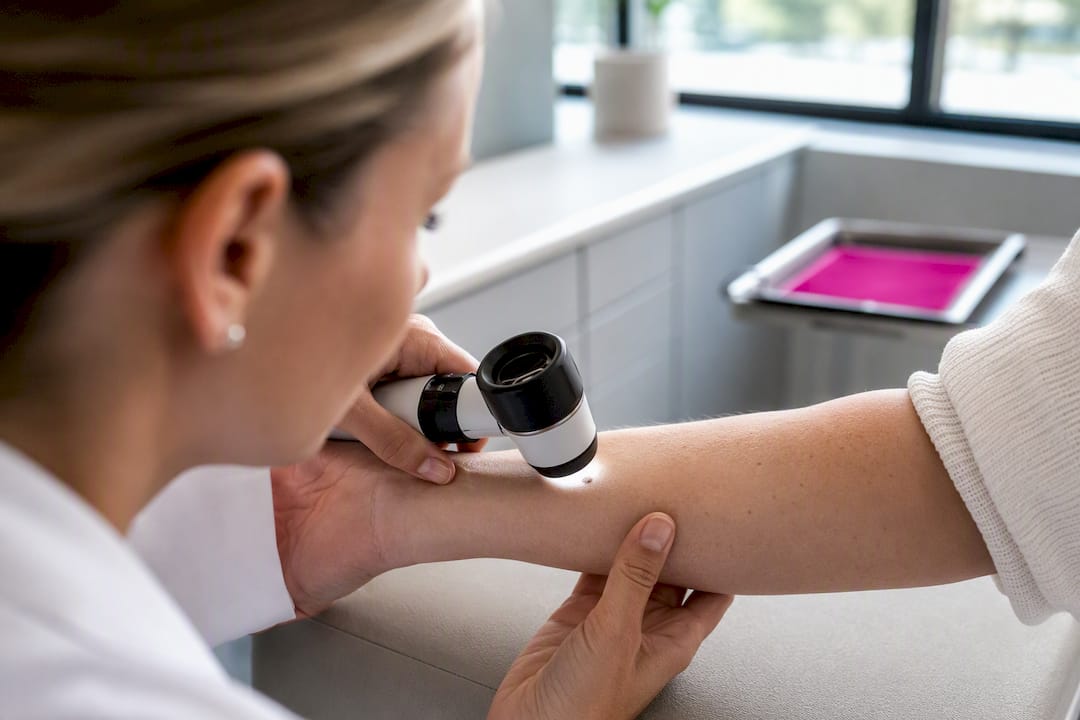
A mole is suspicious when it’s new after age 30, changing in size, shape, or color, or showing any ABCDE feature: Asymmetry, an irregular Border, multiple Colors, a Diameter guidance traditionally points to about the size of a pencil eraser (commonly described as roughly 6mm), but this number is considered less critical than other signs., or Evolving over weeks to months. Bleeding, itching, crusting, or a sore that won’t heal are equally serious. The correct next step is a full evaluation by a board-certified dermatologist. The American Academy of Dermatology notes one in five Americans will develop skin cancer, and A dermatology practice can perform the exam and biopsy needed to confirm or rule it out.
TL;DR:
- Moles that appear after age 30, show recent changes, or fit the ABCDE signs require prompt evaluation by a dermatologist, especially if they bleed, itch, or don’t heal.
- Irregular borders, multiple colors, or sudden darkening in a mole are stronger indicators of concern than size, which has become a less critical factor for melanoma detection.
- The ugly duckling sign and asymmetry are additional red flags, particularly if a mole looks different from all existing moles or breaks your usual pattern.
- Melanomas often develop on palms, soles, under nails, or mucous membranes, making awareness vital for darker-skinned individuals and in non-sun-exposed areas.
- Regular monthly self-checks with photographs and professional skin exams help catch early changes, with biopsy providing the definitive diagnosis.
Table of Contents
- The ABCDEs of Suspicious Mole Signs, Explained
- Ugly Duckling Sign and Other Red Flags Beyond ABCDE
- How to Check Your Skin Every Month and Track Changes
- When to See a Dermatologist and What Happens Next
- Moles in Hard-to-See Spots and What Darker Skin Tones Should Watch For
- Why Trust This Guidance: Rao Dermatology’s Clinical Approach
- A Note on Acting Early Instead of Waiting
- Sources
- FAQ
The ABCDEs of Suspicious Mole Signs, Explained
The ABCDE framework gives you five concrete checkpoints, and each one has a simple test you can run in front of a mirror.
Asymmetry means the two halves of a mole don’t match. Picture drawing a line through the center. A benign mole tends to be a mirror image on each side; a suspicious one looks different from one half to the other.
Border irregularity shows up as scalloped, notched, or blurred edges instead of a smooth, well-defined outline. Think of a coastline versus a circle drawn with a compass.
Color variation is one of the more reliable clues. A single mole showing tan, brown, black, red, white, or blue patches, or one that has simply gotten darker than it used to be, deserves attention even if its shape looks fine.
Diameter guidance traditionally points to about 6mm, roughly the size of a pencil eraser, but this number is the weakest of the five. Dermatologists increasingly flag melanomas smaller than the old diameter cutoff, so a small dark spot that’s changing matters more than a large stable one.
Evolving is arguably the most important letter of all. Any change in size, shape, color, elevation, or symptoms over a matter of weeks or months is a red flag on its own, independent of whether the mole checks any other box.
- A mole with two different colors and a slightly blurred edge
- A spot that was flat six weeks ago and now feels raised
- A mole that used to be uniformly tan and now has a black patch in one corner
Quick fact: the AAD’s ABCDE guidance remains the standard framework dermatologists teach patients for self-checks, precisely because it turns a vague worry into five specific things to look for.
Ugly Duckling Sign and Other Red Flags Beyond ABCDE
Some suspicious moles don’t fit the ABCDE mold cleanly, which is why dermatologists also use the “ugly duckling” sign: a mole that looks noticeably different from every other mole on your body. Most people have a personal pattern to their moles, some oval, some round, mostly one or two shades of brown. A lesion that breaks that pattern, even if it’s technically small and symmetrical, deserves a second look.
Other warning signs worth tracking:
- Persistent itching in a single mole, especially if it’s new
- Bleeding without an obvious injury like a scrape or a shaving cut
- Crusting or oozing that keeps coming back
- A lump or thickened area within an existing mole
- A sore that won’t heal after several weeks
It’s worth noting that pain is not a reliable signal. Many melanomas are painless, so waiting for discomfort before getting checked can delay diagnosis. Non-melanoma skin cancers can look different too. A shiny pink papule or a sore that keeps scabbing over without fully closing can point to basal cell or squamous cell carcinoma rather than melanoma.
Pro Tip: Take a photo of your moles every time you notice something that feels “off,” even if you’re not sure it qualifies as a real change. Comparing two photos side by side is far more reliable than trying to remember what a mole looked like three months ago.
How to Check Your Skin Every Month and Track Changes
A monthly self-exam only works if you do it the same way every time. Here’s a routine that covers the areas people most often skip:
- Start in a well-lit room and check your face, neck, ears, and scalp using a hand mirror or asking a partner to help with the back of your head.
- Work down your chest and torso, then check both sides of your body with your arms raised.
- Examine your back and buttocks using a full-length mirror and a hand mirror together.
- Check your arms, palms, and the spaces between your fingers, then your legs, soles, and the spaces between your toes.
- Inspect your nails for any new dark streaks or bands.
- Don’t skip the genital area and skin folds; melanoma can appear anywhere.
Photograph any mole you’re monitoring under consistent lighting, ideally near a window during daytime, and place a coin or ruler next to it for scale. Date-stamp the image and keep it in a dedicated folder or use a tool like the AAD’s body mole map to log locations systematically. This kind of serial photographic comparison helps clinicians spot evolution that’s easy to miss with memory alone.
Repeat full photo documentation every 4 to 12 weeks depending on your risk level. If a mole changes noticeably before that interval is up, don’t wait. Book an evaluation sooner.
When to See a Dermatologist and What Happens Next
See a dermatologist promptly if you notice a new mole after age 30, a mole that’s changed in size, shape, or color, one that fits the ugly duckling pattern, or any lesion that bleeds, itches persistently, or won’t heal. A persistent lesion that hasn’t resolved within four to eight weeks is a reasonable trigger point many clinicians use to decide whether evaluation is warranted.
A typical visit includes:
- A full-skin exam covering areas you may not check yourself, like your back and scalp
- Dermoscopy, where the dermatologist uses a handheld magnifying device to examine pigment patterns beneath the skin’s surface
- A biopsy if anything looks concerning, followed by pathology review
Biopsy is the only definitive way to diagnose skin cancer. Visual exams and dermoscopy narrow down risk, but a dermatopathologist examining tissue under a microscope is what confirms or rules out a diagnosis. Bring your dated photos to the appointment. They give the dermatologist a timeline that a single snapshot can’t provide, which matters most when the change happened gradually.
Moles in Hard-to-See Spots and What Darker Skin Tones Should Watch For
Melanoma doesn’t stick to sun-exposed skin. Acral and subungual sites, meaning palms, soles, and under the nails, are common locations that get overlooked because people simply don’t look there. A dark, irregular streak or band under a nail, especially one that widens over time or affects a single nail without injury, needs professional evaluation.

Amelanotic melanomas complicate things further because they carry little or no pigment. They can appear pink, red, or the same color as surrounding skin, which means they often skip the “color variation” checkpoint entirely and get mistaken for a pimple, scar, or irritation.
People with darker skin tones face a real blind spot here: melanoma frequently shows up on the palms, soles, nail beds, and mucous membranes rather than sun-exposed areas, and it’s often diagnosed later as a result. Any unexplained pigment change or persistent lesion in these areas deserves a dermatologist’s attention regardless of how much sun exposure you’ve had.
Why Trust This Guidance: Rao Dermatology’s Clinical Approach
This dermatology practice has provided medical and cosmetic skin care across several states for many years. Suspicious lesions get the same clinical workflow every time: a full-skin exam, dermoscopic evaluation, and, when warranted, a biopsy reviewed in collaboration with dermatopathology.
That last step matters. A visual read narrows the possibilities, but tissue analysis is what actually confirms a diagnosis. Patients who want a walkthrough of what to check at home before booking an appointment can start with Rao Dermatology’s at-home skin cancer check guide, which pairs well with the clinic’s skin cancer detection and treatment services for anyone ready to schedule a professional exam.
A Note on Acting Early Instead of Waiting
Most moles people bring in for evaluation turn out to be benign, and that’s the outcome we want. But acting on a change, rather than watching it for another six months, is what actually improves outcomes when something isn’t benign. If a mole has shifted in a way that made you pause long enough to read this article, that instinct is worth listening to. Rao Dermatology’s skin cancer services are there when you’re ready to get a professional opinion instead of guessing.
— Rao Dermatology
This article is general information, not a substitute for advice from a qualified doctor. Consult a qualified healthcare professional about your own circumstances before acting on anything here.
FAQ
What are three warning signs a mole is becoming cancerous?
The three most reliable signs are a change in color (especially new darkness or multiple colors), a change in shape or border regularity, and evolving size or texture over weeks to months. Bleeding or persistent itching are also strong indicators.
What are four early warning signs of melanoma?
Asymmetry, an irregular border, uneven color, and a diameter larger than about 6mm make up four of the five ABCDE warning signs; the fifth, evolving, is often considered the most urgent because any recent change matters regardless of size.
What do early stage cancerous moles look like?
Early melanomas often look like a flat or slightly raised spot with uneven color, ragged edges, and a shape that doesn’t match the rest of your moles. Some are amelanotic and appear pink or skin-colored instead of dark, which makes them easy to miss.
What counts as a suspicious mole?
A mole counts as suspicious if it’s new after age 30, doesn’t match your other moles (the ugly duckling sign), shows any ABCDE feature, or bleeds, itches, crusts, or fails to heal. Any of these on their own is reason enough for a dermatologist to take a look.
Recommended
Filed under: